
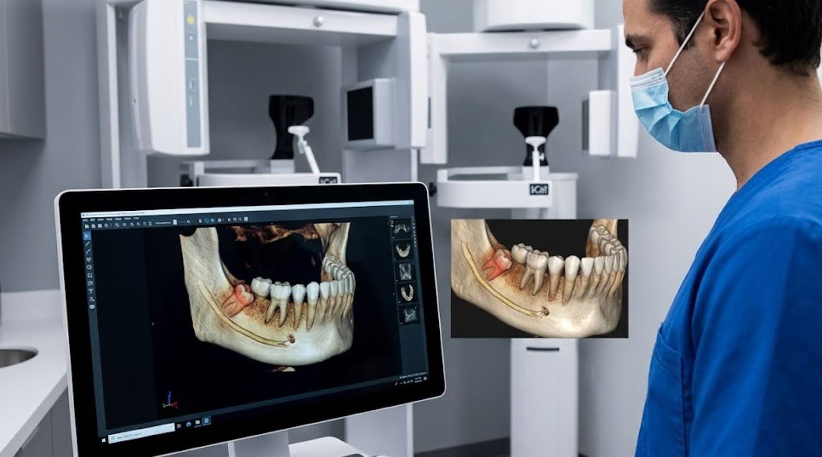
Wisdom tooth extraction is a routine procedure but carries measurable risks when anatomical relationships are not fully known. iCat 3D Imaging has changed how oral surgeons plan and perform these extractions by providing detailed three-dimensional information that improves surgical precision and patient safety. This article explains how iCat 3D Imaging enhances safety, reviews the technology’s clinical advantages, and outlines why Siouxland Oral Surgery is a preferred provider in Sioux Falls. Readers will learn how the system works, how it compares with traditional imaging, and the practical impact on patient outcomes.
What is iCat 3D Imaging and How Does It Improve Oral Surgery Imaging?
iCat 3D Imaging is a cone beam computed tomography (CBCT) system that produces volumetric, three-dimensional views of the teeth, jaws, and adjacent tissues. By visualising complex anatomical relationships, surgeons can plan extractions with greater accuracy. The detailed images allow assessment of tooth position and proximity to critical structures such as the inferior alveolar nerve and the maxillary sinuses, supporting more informed surgical decisions and targeted treatment strategies in oral surgery sioux falls sd.
Although iCat 3D Imaging provides clear advantages in many cases, clinical research continues to refine the specific indications for its routine use, particularly with respect to preventing nerve injury in high‑risk wisdom tooth removals.
CBCT for Wisdom Teeth: Nerve Damage & Safety
There is ongoing discussion about whether a CBCT scan should be performed before surgical removal of wisdom teeth. A prospective, randomized, controlled multicentre trial sought to confirm non‑inferiority for inferior alveolar nerve damage when patients were diagnosed by panoramic radiography compared with CBCT. The findings indicate that CBCT scans should generally be reserved for high‑risk wisdom tooth removals.
Necessity of 3D visualization for the removal of lower wisdom teeth: required sample size to prove non-inferiority of panoramic radiography compared to CBCT, R Schulze, 2012
Benefits of iCat 3D Imaging
iCat 3D Imaging delivers several clinical benefits that contribute directly to safer, more effective wisdom tooth extractions. The principal advantages include:
- Enhanced Accuracy: The three-dimensional images provide a comprehensive view of the dental anatomy, allowing for precise surgical planning.
- Reduced Surgical Surprises: Surgeons can identify potential complications before the procedure, minimizing unexpected challenges during surgery.
- Lower Radiation Exposure: Compared to traditional imaging methods, iCat 3D Imaging typically requires less radiation, making it a safer option for patients.
| Benefit | Description | Impact Level |
|---|---|---|
| Enhanced Accuracy | Provides detailed 3D views of oral structures | High |
| Reduced Surgical Surprises | Identifies potential complications in advance | High |
| Lower Radiation Exposure | Uses less radiation than traditional X-rays | Medium |
Taken together, these advantages reduce intraoperative risk and support more predictable recoveries for patients undergoing wisdom tooth extractions.
Understanding Cone Beam Computed Tomography Technology
Cone beam computed tomography (CBCT) is the core technology behind iCat 3D Imaging. Unlike conventional two‑dimensional X‑rays, CBCT acquires image data from multiple angles and reconstructs a three‑dimensional model of the patient’s anatomy. This volumetric representation yields a more accurate depiction of jaw morphology and bone density, which is essential for planning complex extractions and reducing procedural uncertainty.
How Does iCat 3D Imaging Reduce Radiation Exposure Compared to Traditional X-Rays?
iCat 3D Imaging can lower cumulative radiation exposure because a single volumetric scan often provides the diagnostic information that would otherwise require multiple two‑dimensional images. Reducing the number of separate exposures is especially relevant for younger patients and for those who may need repeated imaging over time.
Benefits of Reduced Radiation Dental Scans for Patient Safety
The safety benefits of reduced radiation exposure with iCat 3D Imaging are meaningful. Key points include:
- Health Benefits: Lower radiation doses reduce potential long‑term risks associated with ionising radiation.
- Impact on Patient Comfort: Patients typically appreciate imaging options that minimise exposure while providing clear diagnostic information.
- Long-Term Safety Considerations: Minimising radiation is important for patients who may require multiple dental procedures over their lifetime.
In What Ways Does iCat 3D Imaging Enhance Safety During Complex Third Molar Surgery?
iCat 3D Imaging enhances safety in complex third molar surgery by clearly delineating the relationship between the third molar and adjacent vital structures. Surgeons use these images to plan incisions and instrument trajectories that avoid nerves and major vessels, reducing the risk of nerve injury and excessive bleeding. Accurate preoperative imaging supports a controlled surgical approach and can contribute to faster, less complicated recoveries.
Precise Surgical Planning Enabled by 3D Imaging Techniques
The precision afforded by iCat 3D Imaging enables meticulous surgical planning. Surgeons can use 3D data to design patient‑specific surgical guides and to position instruments with greater accuracy during extraction. This level of preparation shortens operative time, reduces the likelihood of intraoperative complications, and supports improved clinical outcomes.
Combining advanced imaging with virtual surgical planning illustrates how modern tools increase both precision and safety in complex oral procedures.
3D Virtual Surgical Planning for Precision & Safety
Virtual surgical planning (VSP) and 3D printing have advanced surgical precision and customisation. VSP enables three‑dimensional visualisation of the operative site and real‑time adjustments, which can reduce preoperative stress by shortening planning time. 3D printing complements VSP by producing anatomical models and surgical guides that improve the predictability of outcomes, despite higher initial setup and material costs. The integration of VSP and 3D printing offers practical solutions that address limitations of traditional methods in orthognathic surgery.
Redefining precision and efficiency in orthognathic surgery through virtual surgical planning and 3D printing: a narrative review, SG Kim, 2023
Monitoring Recovery and Detecting Complications Early
Postoperative monitoring is essential to ensure uncomplicated recovery after wisdom tooth extraction. Follow‑up CBCT scans with iCat can document healing and reveal early signs of complications, such as infection or atypical bone healing. Early detection permits timely intervention, which improves recovery trajectories and patient comfort.
Patient Testimonials on Safety and Comfort Improvements
Patients who have had extractions planned with iCat 3D Imaging commonly report greater confidence and satisfaction. Many note appreciation for thorough preoperative assessment and reduced anxiety from knowing the surgical team used advanced imaging to guide care. These testimonials reinforce the value of integrating precise imaging into oral surgery practice.
Why Choose Siouxland Oral Surgery for Wisdom Teeth Extractions with Advanced 3D Imaging?
Siouxland Oral Surgery is a leading option in Sioux Falls because the practice integrates advanced imaging like iCat 3D into a patient‑centred model of care. Each patient receives personalised evaluation and a treatment plan tailored to their anatomy and clinical needs. With a team of experienced oral surgeons, Siouxland prioritises safety, comfort and predictable outcomes for patients undergoing wisdom tooth removal.
By choosing Siouxland Oral & Maxillofacial Surgery, patients can expect a comprehensive approach that leverages current imaging advances to improve procedural safety and clinical results.
Frequently Asked Questions
What are the potential risks of wisdom teeth extractions without advanced imaging?
Extraction without advanced imaging can increase the likelihood of complications because two‑dimensional techniques may not fully reveal the tooth’s relationship to nearby nerves and other structures. Potential risks include nerve injury, excessive bleeding and incomplete removal. Advanced three‑dimensional imaging improves preoperative planning and helps minimise these risks.
How does iCat 3D Imaging compare to traditional X-rays in terms of patient comfort?
iCat 3D Imaging often improves patient comfort relative to traditional X‑rays. A single volumetric scan typically captures the necessary diagnostic information more quickly than multiple two‑dimensional exposures, reducing chair time. Lower cumulative radiation exposure also helps address safety concerns, particularly for younger patients.
Can iCat 3D Imaging be used for other dental procedures besides wisdom teeth extractions?
Yes. iCat 3D Imaging is useful for implant planning, orthodontic assessment and a range of complex oral surgeries. The detailed three‑dimensional views allow clinicians to visualise anatomical structures accurately, enabling more precise planning and execution across multiple treatment types and improving overall patient outcomes.
What should patients expect during the iCat 3D Imaging process?
The iCat scan is a quick, non‑invasive procedure that usually takes only a few minutes. Patients are positioned comfortably in the scanner and asked to remain still while the device acquires images from different angles. The process is painless, and most patients can resume normal activities immediately afterward.
How does iCat 3D Imaging contribute to post-operative care?
iCat 3D Imaging supports post‑operative care by enabling follow‑up scans to monitor healing and to identify complications such as infection or abnormal bone repair early. Prompt identification of issues allows clinicians to intervene sooner, which can streamline recovery and enhance long‑term outcomes.
Are there any age restrictions for using iCat 3D Imaging?
There are no absolute age restrictions for iCat 3D Imaging. The decision to use CBCT is based on clinical need and individual health considerations. It is particularly advantageous for younger patients who may require wisdom tooth extraction, as it can reduce overall radiation exposure compared with multiple conventional X‑rays. Dental professionals assess each case to determine appropriateness.
Conclusion
iCat 3D Imaging enhances safety and comfort in wisdom tooth extractions by improving anatomical visualisation and reducing cumulative radiation exposure. This technology enables oral surgeons to plan procedures with greater precision, thereby minimising risk and supporting more predictable outcomes. Patients who choose Siouxland Oral Surgery benefit from advanced imaging integrated into personalised care. To learn how iCat 3D Imaging can inform your treatment, book an appointment with the practice.